Digital X-Ray
Jupiter, FL
A great deal of what happens inside your mouth is invisible to the naked eye. Cavities developing between teeth, bone loss occurring beneath the gumline, infections forming at the tip of a root, and impacted teeth positioned deep within the jaw all exist in spaces that even the most thorough visual examination cannot access. Dental X-rays have long been the tool that bridges that gap, and the shift to digital technology over the past two decades has made that tool faster, safer, and far more informative than its film-based predecessor. Understanding how digital X-rays work and what they reveal helps patients at Roy C. Blake III, DDS, MSD, Maxillofacial Prosthodontist appreciate why they are a routine and important part of dental care. What Digital X-Rays Actually AreDigital X-rays, like traditional film X-rays, use a small amount of radiation to produce images of the teeth, roots, and surrounding bone. What differs is how those images are captured and processed. Instead of film that must be developed in a darkroom using chemical solutions, digital X-rays use an electronic sensor placed inside the mouth. The sensor captures the image when the X-ray beam passes through the teeth and bone, and that image appears on a chairside monitor within seconds. Two types of sensors are used in digital dental imaging. Direct digital sensors are hard, wired devices connected to the computer that capture and transmit images instantly. Phosphor plate sensors, sometimes called indirect digital sensors, are thin and flexible like traditional film and are scanned after exposure to produce the digital image. Both approaches offer significant advantages over conventional film, with direct sensors providing the fastest turnaround and phosphor plates offering greater flexibility in positioning for certain patients. How the Images Are ProducedWhen the X-ray beam passes through the structures of the mouth, different tissues absorb the radiation at different rates. Dense structures like enamel and bone block more of the beam, appearing lighter on the image. Less dense structures like soft tissue and areas of decay absorb less, appearing darker. This contrast is what allows the dentist to identify problems that would otherwise be hidden. With digital sensors, those contrast values are captured electronically and rendered as a high-resolution image on the monitor. The image can be enlarged, brightened, adjusted for contrast, and annotated, giving the provider tools to identify subtle changes that might be missed on a smaller or lower-resolution film image. The ability to manipulate the image without losing quality is one of the most meaningful practical advantages of digital X-rays for diagnostic purposes. Types of Digital X-Rays Used in DentistryDifferent diagnostic needs call for different types of X-ray images. Each type captures a specific area or perspective of the oral structures.
Digital X-Rays vs. Traditional FilmThe differences between digital and film X-rays are meaningful in both the patient experience and the quality of diagnostic information produced. Film required patients to hold a sharp-edged film packet in the mouth while it was developed off-site or in an in-office darkroom, a process that took considerably longer and involved chemical developers that required careful handling and disposal. Digital sensors are thinner and more comfortable than film packets in many cases, and the images are available immediately. There is no waiting, no chemical processing, and no risk of a film being accidentally exposed or lost. For patients, this means shorter appointments and faster answers. For providers, it means more time focused on diagnosis and less on administrative handling of film. Radiation exposure is also meaningfully lower with digital X-rays. Depending on the system used, digital sensors require significantly less radiation than conventional film to produce a comparable or superior image, making an already low-risk procedure even safer. How Digital X-Rays Support Better DiagnosisOne of the most important advantages of digital X-rays is how they support communication between the dentist and the patient. When an image is displayed on a large chairside monitor at the moment of capture, the dentist can walk the patient through exactly what they are seeing. A cavity that is just beginning to form, a hairline fracture in a root, or a slight reduction in bone density can be shown and explained in real time rather than described in abstract terms. This visibility also supports more accurate documentation over time. Digital images are stored in the patient's electronic record and can be compared directly to images taken at previous visits. Changes in bone levels, the progression of a cavity, or the healing of a treated area are all visible in side-by-side comparison, giving both the provider and the patient a clearer picture of how oral health is evolving. Are Digital X-Rays Safe?Radiation exposure from dental X-rays is among the lowest of any medical imaging procedure. The amount of radiation in a full set of dental bitewing X-rays is comparable to the natural background radiation a person receives from the environment over the course of a day or two. Digital X-rays reduce that already minimal exposure further by requiring less radiation to produce a clear image. Protective measures, including a lead apron placed over the patient's chest and lap during imaging, further reduce incidental exposure to other parts of the body. Thyroid collars are also available and may be used for patients who prefer additional protection. These precautions, combined with digital technology's reduced radiation requirements, make routine dental X-rays a very low-risk part of preventive care. Experience the Difference at Our Office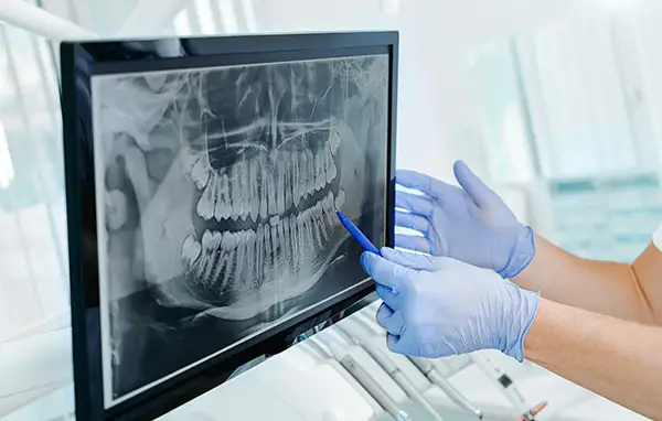
At Roy C. Blake III, DDS, MSD, Maxillofacial Prosthodontist, we use digital X-ray dental technology as a core part of every comprehensive dental evaluation. Our dental team is happy to walk you through each image and explain exactly what we are looking for and what we find. To schedule an appointment or ask questions about our imaging process, call us at (561) 296-3399 today. |
"Dr. Blake is always extremely professional, meticulous, accommodating, and at the same time, affable and sincerely interested in his patient’s well-being and concerns. The staff is at all times very courteous and professional. Their constant friendly and pleasant attitudes ensure that at every visit, the patient’s experience is always relaxing and positive." -John |